UHRF1 Protein Overexpression and Gene Silencing in Adult T-Cell Leukemia/Lymphoma
Researchers analyzed tissue samples from 102 individuals and found that UHRF1 protein abundance is significantly elevated in adult T-cell leukemia/lymphoma patients. Furthermore, experimental silencing of this factor in malignant T-cell lines successfully reduced cell growth rates and triggered cell death.
Reviewed by Matheus GrangeiroUpdated on
- Peer reviewed
- Yes
- Sample size
- 102
Key numbers
72patients
ATLL patient cohort size
30patients
Reactive lymphadenopathy control group size
26.562median optical density
UHRF1 expression optical density in primary ATLL samples
1.306median optical density
UHRF1 expression optical density in control group
9.914percent
Positive pixel percentage area in ATLL group
0.574percent
Positive pixel percentage area in control group
2.397hazard ratio
Overall survival hazard ratio for UHRF1 expression
1.959hazard ratio
Progression-free survival hazard ratio for UHRF1 expression
Evidence level: Retrospective clinical cohort plus preliminary in vitro experiments
Why it matters: Patients diagnosed with adult T-cell leukemia/lymphoma (ATLL) face poor survival and progression outcomes, and the specific contribution of susceptibility factors such as UHRF1 to ATLL pathogenesis had remained poorly defined. Identifying an independently adverse prognostic biomarker could help refine risk stratification, while preliminary functional evidence may point to future therapeutic targets.
Evaluating UHRF1 expression and outcomes in ATLL Ying Lin, Rong-Dong Zhang, Xin-Yu Xu, Huang-zhou Jiang, Ren-Li Chen and Jian-Zhen Shen evaluated UHRF1 protein abundance by immunohistochemistry in tumour tissues from 72 patients with ATLL and 30 patients with reactive lymphadenopathy. The samples came from patients diagnosed or previously diagnosed between January 2018 and January 2025 at Ningde Municipal Hospital and Fujian Medical University Union Hospital.
The ATLL cohort had a mean age of 57.5 years (standard deviation, 12.4 years) and comprised 37 men and 35 women. ATLL is an aggressive malignancy characterized by clonal expansion of mature CD4-positive T cells associated with persistent human T-cell leukemia virus type I (HTLV-1) infection.
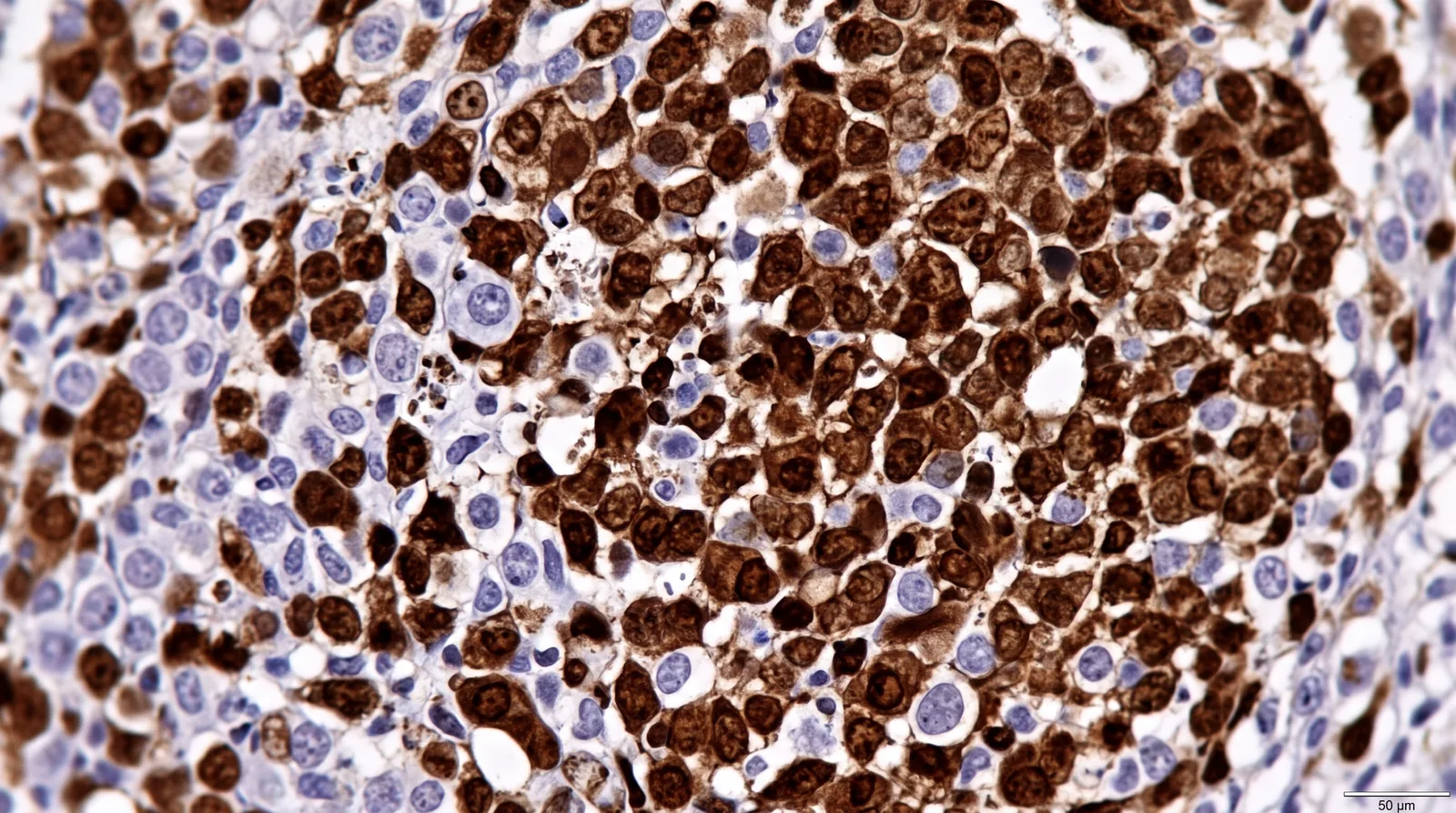
Two experienced pathologists independently scored UHRF1 staining while blinded to clinical outcomes. UHRF1 expression was substantially higher in ATLL tissues than in controls: the median optical density was 26.562 versus 1.306, and the percentage of positive stained area was 9.914% versus 0.574%; both differences were statistically significant (P < 0.001).
Survival associations and prognostic modelling During a median follow-up of 10.5 months, 56 patients experienced disease progression and 43 died; no patients were lost to follow-up. The researchers used univariate and multivariate Cox proportional-hazards regression to assess overall survival (OS) and progression-free survival (PFS).
After adjustment for clinicopathological and laboratory variables, high UHRF1 expression remained an independent adverse prognostic factor. It was associated with poorer OS (hazard ratio [HR], 2.397; 95% confidence interval [CI], 1.108–5.184; P = 0.026) and poorer PFS (HR, 1.959; 95% CI, 1.025–3.744; P = 0.042).
High UHRF1 expression was also associated with adverse disease features, including acute or lymphoma-type ATLL, B symptoms, bone-marrow invasion, higher LDH, beta-2 microglobulin, CRP, Ki-67 and HTLV-1 viral load. These are associations in a retrospective cohort, not evidence that UHRF1 causes these clinical features.
Testing UHRF1 silencing in ATLL cell lines In preliminary experiments, the researchers transduced HuT-102 and MT-4 ATLL-derived cell lines with lentiviral vectors carrying short hairpin RNAs (shRNAs) targeting UHRF1. Cells were transduced in suspension culture with polybrene and selected with puromycin; among three tested target sequences, KD2 showed the greatest knockdown efficiency and was used for subsequent functional assays.
UHRF1 silencing significantly suppressed proliferation in both cell lines. In the CCK-8 assay, conducted over a five-day culture period, absorbance at 450 nm was significantly lower in the knockdown group than in the scrambled-shRNA control from day 2 onward.
UHRF1 knockdown also induced apoptosis. In HuT-102 cells, it increased both early and late apoptotic cell fractions; in MT-4 cells, it markedly increased early apoptosis and reduced the viable-cell fraction. Flow cytometry using Annexin V-FITC and propidium iodide staining supported these observations.
Unresolved signalling mechanisms and protein interactions The study also examined whether UHRF1 interacts with the AMPK signalling pathway. Immunofluorescence showed partial subcellular overlap between UHRF1 and AMPK alpha 1 (PRKAA1), but this finding alone cannot establish a physical interaction.
Co-immunoprecipitation did not detect a stable interaction between UHRF1 and either AMPK or the catalytic subunit of protein phosphatase 2A, PPP2CA. In addition, UHRF1 knockdown did not significantly alter LKB1, PPP2R4 or the ratio of phosphorylated AMPK at Thr172 to total AMPK.
These negative and inconclusive results mean that the intracellular pathway through which UHRF1 may promote ATLL cell growth remains uncharacterized. The authors themselves state that whether UHRF1 intersects with AMPK signalling remains to be determined.
What the study does not show It may seem that UHRF1 is a validated therapeutic target for ATLL. In fact: the functional evidence comes from two cell lines and preliminary shRNA experiments; no patient treatment, animal model or clinical intervention was tested.
The prognostic association comes from a retrospective, single-country cohort of 72 patients and requires external validation in larger and more diverse populations.
The lack of a detected UHRF1–AMPK or UHRF1–PPP2CA interaction does not prove that no interaction exists; it indicates that these specific co-immunoprecipitation assays did not detect one under the tested conditions.
The mechanisms linking UHRF1 to proliferation, apoptosis and cell-cycle changes remain unresolved and require further biochemical and in vivo validation.
Verdict: In a retrospective cohort of 72 patients with ATLL, elevated UHRF1 expression was independently associated with poorer overall and progression-free survival. Preliminary experiments in HuT-102 and MT-4 cells showed that UHRF1 knockdown reduced proliferation and increased apoptosis, but the downstream mechanism remains unmapped and the findings require validation in larger cohorts and in vivo models.
Source: PLOS ONE. The original is licensed CC BY 4.0. This text is an AI-assisted adaptation (summarized, simplified and translated) and may differ from the original.